

OUR PRODUCTS
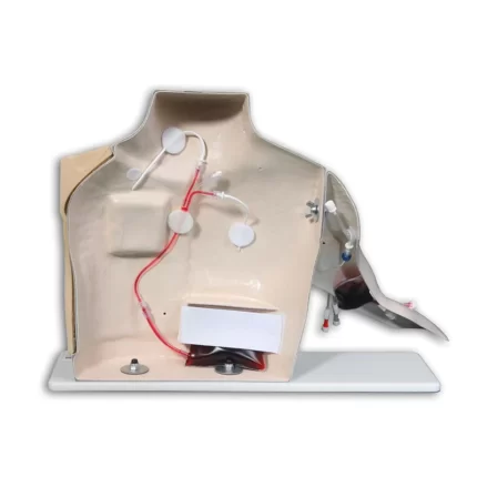
Chester Chest™ Vascular Access Simulator With Port Access Arm
Chester Chest™ has been an industry standard since 1986 for the teaching of central line care. The right chest area of Chester Chest™ has a 9.6Fr tunneled central catheter that is visible up to the clavicle. The Dacron cuff on this catheter is also discernable. The external jugular vein is slightly raised with an opening to attach your own triple lumen catheter and there is also an opening in the upper chest area for placement of a subclavian catheter. These optional catheters are available from VATA, or if you prefer, you can send us the catheters your institution uses and we will install them at no charge.
Included with this model is a real port that makes it possible to simulate accessing the following IVAD placements: normal, “tipping”, “wandering”, or deeply placed in the left chest area. Successful access is confirmed by a blood return. Fluid can be infused and blood withdrawn from all the lines.
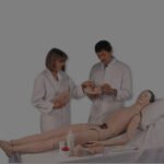
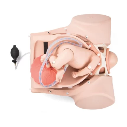
3B Birthing Simulator PRO
The 3B Scientific® Birthing Simulator P90 PRO has been developed for the skill training in normal deliveries, in complicated deliveries and in obstetric emergencies. Obstetric simulation has proven successful to enhance the training of delivery skills, following of protocols and reaction in emergency situation.
Assessment and manipulation of fetal positions:
Birthing complications are in general less likely when an abnormal fetal position or presentation can be detected before the labor process starts. Using obstetric simulation, trainees will learn how to detect abnormal positions and presentations, and how to use manual techniques to assist the birthing process. Training of manual birthing maneuvers (like Leopold, or Pinard’s) must be trained so that correct measures are applied during complicated deliveries. Furthermore, the trainee will learn when to apply obstetric emergency interventions (like a cesarean section).
Replacement Tissue for Epidural and Lumbar Puncture
This Replacement Tissue for Obese Lumbar Epidural and Lumbar Puncture is designed to be used with item BPLP210. The obese spinal insert provides more adipose tissue disallowing the palpation of the spinal processes. This module is superb for needle access as well as the placement of catheters.
Pediatric Head Simulator
This pediatric injectable head model provides realistic sensation and response. The lifelike vinyl skin actually rolls as you palpate to allow location of the vein. The synthetic rubber tubing for the veins was carefully selected to provide a lifelike simulation of vein size, and feeling of puncture and palpation for practicing venipuncture. The temporal vein of the Life/form® Pediatric Head is easily accessible for IV infusions. Practice in the jugular vein is equally realistic. The neck is made of soft, flexible foam to provide a realistic feel of palpation and puncture. Includes a Life/form® Head with skin and veins, fluid supply bag, two different gauge winged infusion needles, one pint Life/form® blood, teaching guide, and hard carrying case.
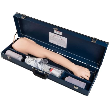
Advanced Venipuncture & Injection Arm
Life/form® Advanced Venipuncture and Injection Arm
This revolutionary Life/form® Advanced Venipuncture and Injection Arm, white skin, provides complete venous access for IV therapy and phlebotomy, plus sites for intramuscular and intradermal injections. An extensive 8-line vascular system allows students to practice venipuncture at all primary and secondary locations, including starting IVs and introducing Over the Needle IV Catheters.
The venous system simplifies setup with only one external fluid bag supplying artificial blood to all veins simultaneously. The dorsal surface of the hand includes injectable metacarpal, digital, and thumb veins. The antecubital fossa includes the median cephalic, median basilic, and median cubital veins.
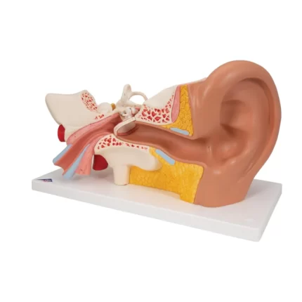
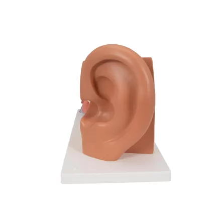
Giant Ear Model, 3x Life Size, 4-part
Giant Ear Model, 3x Life Size, 4-part - Includes 3B Smart Anatomy
This high quality model of the human ear represents outer, middle and inner ear. The detailed human ear model has removable eardrum with hammer, anvil and stirrup as well as 2-part labyrinth with cochlea and auditory/balance nerve. Ear on base for easy display in a classroom or doctor's office. This ear model is a great way to teach and study the anatomy of the human ear!Gen II Ultrasound Central Line Training Model
SOMSO Transparent Human Skull with Brain Model
SOMSO Transparent Human Skull with Brain Model
Brain separates into 8 parts: frontal and parietal lobes (2), temporal and occipital lobes (2), medulla (2), cerebellum (2). Transparent Skull: removable vault, lower jaw movable, separates into 3 parts.Cleaning and disinfecting your SOMSO Model: Spray a rapid and effective water based disinfectant for use on surfaces and wipe with a soft cloth. The disinfectant spray must be pH neutral, alcohol-free, aldehyde free, fragrance-free and nonflammable.
About Alpha Century Simulations
Established in 2015, Alpha Century Simulations has become one of the top companies in Nigeria supplying medical simulators and anatomical models to the Educational Market.
Health care demands are our single focus and they motivate us to carry the best line of products and to invest in training and customer care……

WHAT WE OFFER


