


Head and Neck Musculature Model, 5-part
Head and Neck Musculature Model, 5-part - Includes 3B Smart Anatomy
Representation of the superficial musculature and deep muscles, nerves and vessels. The head and neck musculature model can be dissected into skull cap and 3-part brain. This high quality model delivered on removable baseboard for easy use in a classroom or doctor's office. All important anatomical structures of the human head are detailed.
Head Musculature Model with Blood Vessels


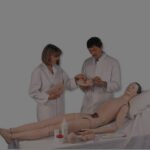
Heart and Lung Sounds Adult Torso
Heart and Lung Sounds Adult Torso
The amazing technology that simulates the various heart and breath sounds is hidden within the stethoscope bell itself. There are no bulky boxes or transmitters, making it easy to use and realistic. Simply press the bell against the simulator’s skin at the correct anterior or posterior auscultatory site and listen to the corresponding sound. The VS100/VS105 include a reference key card that indicates the auscultatory sites locations and the sound types available. Additionally, the package includes a set of optional loud speakers useful when teaching an audience.
Features
Hear the appropriate heart or lung sound as bell of stethoscope is moved across the front and back of the torso
Full size adult torso with palpable anatomic landmarks
Sensor network hidden beneath the skin
Includes our Virtual Stethoscope® with multiple heart and lung sounds
Instruction manual
An external speaker plugs into the Virtual Stethoscope so a classroom can hear what the student hears
Carrying bag

Lumbar Disc Herniated, 2x Life-size


Lung Model with Larynx and Heart, 7 Part
Lung Model with Larynx and Heart, 7 Part - Includes 3B Smart Anatomy
The lung model with larynx is first class. The high quality lung model contains the following removable parts for added anatomical detail:- 2-part larynx
- Trachea with bronchial tree
- 2-part heart
- Subclavian artery and vein
- Vena cava
- Aorta
- Pulmonary artery
- Esophagus
- 2-part lung (front halves removable)
- Diaphragm

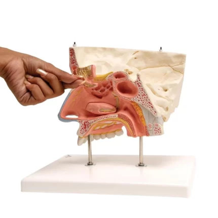
Nose Model with Paranasal Sinuses, 5 part
Nose Model with Paranasal Sinuses, 5 part - Includes 3B Smart Anatomy
This nose model illustrates the structure of the nose with the paranasal sinuses in the upper right half of a face in 1.5-fold enlargement. The following structures can be seen from the outside of the nose with paranasal sinuses, differentiated by color (also visible through the removable transparent skin):- The outer nasal cartilages
- The nasal cavity, maxillary, frontal and ethmoid sinuses
- The opened maxillary sinus when the zygomatic arch is removed
- The nasal cavity, lined with mucosa, with the nasal conchae (removable)
- The arteries of the mucous membrane
- The olfactory nerves
- The innervation of the lateral wall of the nasal cavity, the nasal conchae and the roof of mouth (palate)
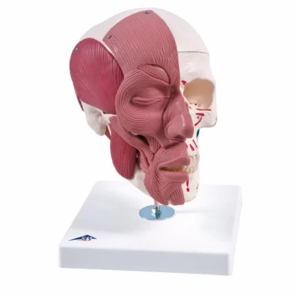
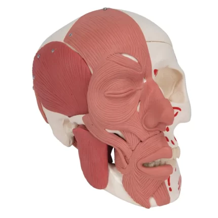
Skull Model with Facial Muscles
The face and mastication muscles are illustrated on the right half of this skull model. The face musculature can easily and precisely be differentiated from the mastication musculature by using two colours. On the left half the muscle origins and insertions are marked with colours (origin: red, insertion: blue). The jaw is movable and due to the flexible musculature the rudimentary chewing motion can be demonstrated. Cranium and m. masseter are detachable.